|
|
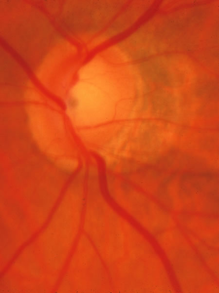
| Fig. 8. Tilted disc, with vessel trunk beneath a nasal ledge, a sloping disc surface on the temporal side, and a peripapillary crescent (β zone) the edge of which envelops the upper and lower poles. Although difficult to judge at times in tilted discs, the neuroretinal tissue is thinned at the poles of the disc in a manner typical of glaucoma. |