|
|
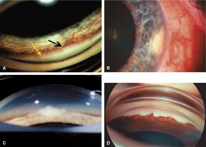
| Fig. 14 Mystery angles (answers in body of text in last paragraph). A. Goniophotograph captured through a Koeppe lens during an examination under anesthesia (EUA) of a 4-year-old with suspicious optic discs and borderline intraocular pressures (IOPs). The iris sweeps from the recess all the way up to a prominent Schwalbe's line. The appearance of the iris is almost transparent or diaphanous; it is not opaque because the majority of the underlying angle structures are visualized. The black arrow indicates iris processes that sweep up into the angle; these are not peripheral anterior synechiae. The red arrow points to the ciliary body band, the scleral spur is poorly seen. The yellow arrow indicates a prominent Schwalbe's line. The fellow eye has the same appearance. What is your diagnosis? B. Slit-lamp photograph of a limbal lesion. The lesion appears confined to the cornea and the IOP is normal. Treatment included aggressive cryotherapy of the area. C. The goniophotograph of (B) reveals the lesion has invaded the chamber angle. A white mass is seen in the angle. What is your diagnosis? D. Goniophotograph obtained through a Koeppe lens immediately prior to trabeculectomy for uncontrolled glaucoma. The angle process is bilateral. The patient is 54-year-old female with a 30-year history of initially open-angle glaucoma. The angle originally appeared open; however, over the past 10 years has developed the captured appearance. Note the difference in the characteristics of the iris. No angle structures are seen underneath the iris. It is totally opaque, not diaphanous as in figure a. These peripheral anterior synechiae (PAS) bridge the angle far onto the cornea, a highly abnormal position for the iris except for a few conditions. What is your differential and diagnosis? If a unilateral process, iridocorneal endothelial syndromes (ICE) would be first. This process is bilateral. The corneal endothelium demonstrates various areas of endothelial plaques, vesicles, and striae. (Figure continues.) |