|
|
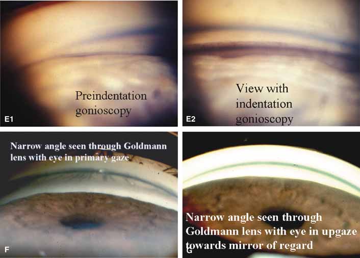
| Fig. 13b Continued. E–1. View into the chamber recess in a narrow angled eye. Figure e–1 is the preindentation appearance. Only a small amount of pigment is seen and landmarks are lost. E–2. The angle structures are easy to see with indentation. As the iris is temporarily pushed back, the heavy pigmentation of the meshwork is seen and the irregular insertion of the iris onto the ciliary body is noted. This is angle closure at an early stage. The PAS are starting to develop deep in the recess. Indentation gonioscopy permitted the correct diagnosis at an early stage and this patient did well with a peripheral iridotomy. F. View of narrow angle as seen through a Goldmann lens. No angle structures are visualized. How would you grade this angle? Shaffer grade 2 to 3, obviously narrow. The Spaeth system requires a grade of the apparent insertion of the iris before and after indentation. The apparent insertion is designated by parentheses. Preliminary Spaeth = (A)20b3+. G. View of narrow angle as seen through Goldmann lens with patient looking in direction of mirror. Sometimes this maneuver will bring the recess into view. The angle is so narrow in this case that it does not help. (Figure continues.) |