|
|
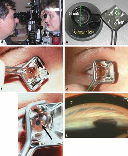
| Fig. 12b Continued. G. Both the examiner and patient are comfortable. The examiner rests his or her elbow on the slit-lamp base for stability while holding on to the gonioprism. The patient is comfortable at the slit lamp with their eye lined up perfectly for indirect gonioscopic evaluation. H. Difference in contact size between Goldmann and Zeiss lenses. The smaller contact area of the Zeiss lens causes less angle distortion especially when trying to view a narrow angle. The smaller area negates the need for a viscous bridge. I. The Zeiss lens inserted with the diamond orientation causes excessive contact with the lids and increases pain. Some examiners prefer this configuration. This is related to the variable handle designs and holding patterns adopted around the world. J. The square orientation is preferred. The lens causes less pain in this position and all four mirrors are then evaluated. K. This incomplete capillary bridge (black arrow) is easily seen in this picture. When learning Zeiss gonioscopy, it is imperative to evenly distribute the tear film, which implies the pressure across the cornea is roughly equal. When learning Zeiss gonioscopy, the examiner should rest their fourth and fifth digits on the patient's cheek for stabilization and orientation, look directly at the patient's eye when starting to insert the prism, and after globe contact, look through the biomicroscope. L. Excessive or uneven pressure on the cornea will cause corneal striae. Initially this is common and the examiner must anticipate this and adjust the force on the cornea. (Figure continues.) |