|
|
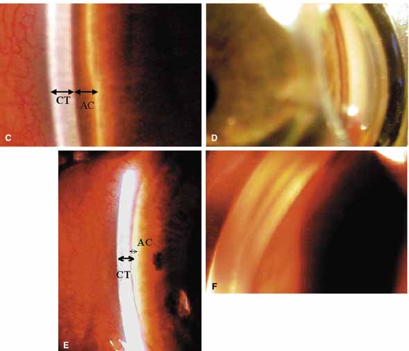
| Fig. 9b Continued. C. The van Herick test is an estimate of angle width. The slit lamp beam is aligned at the limbus and peripheral iris at approximately 60 degrees. The corneal thickness (CT), is compared to the anterior chamber depth (ACD). The scale and corollary to the Shaffer system is shown in Table 2 In this example, a van Herick grade 4 is noted. D. This is the goniophotograph of the actual angle from the grade 4 van Herick in (C). The exact location of the spur, the amount of pigment in the meshwork, where the iris inserts into the angle are now known. The van Herick estimate was popular during the reign of office based Koeppe gonioscopy, which was burdensome. Modern-day indirect gonioscopy allows a rapid assessment of the angle while the van Herick remains an estimate. E. Another example with peripheral iris closer to limbal cornea. This is a van Herick grade 2. F. In this case the iris appears close to the cornea not because of pupil block and iris bowing, but because of a high insertion of the iris into the ciliary body band. The clinical point is when the van Herick is narrow; it is impossible to know if it is due to forward bowing of the iris as in pupil block or a high insertion of the iris as in this case. |