|
|
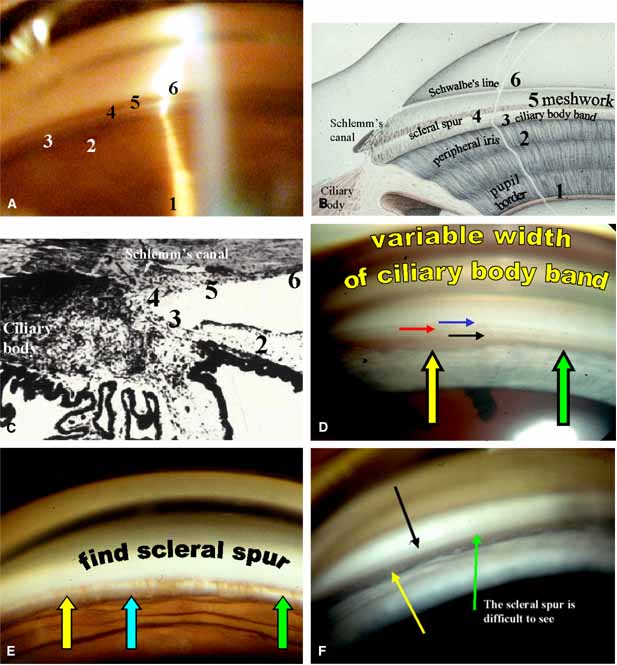
| Fig. 3 The normal angle. A. Can you identify structures 3, 4, 5, and 6? (correlate with B and C). B. This drawing identifies the six key structures to evaluate during gonioscopy: (1) pupil border; (2) peripheral iris; (3) ciliary body band; (4) scleral spur; (5) trabecular meshwork; and (6) Schwalbe's line. C. It is useful to correlate the goniophotograph with the histology of the angle. It is obvious the depth of the angle recess is partly dependent on where the iris inserts onto the ciliary body band. D. Ciliary body band width varies considerably. This goniophotograph is a developmental abnormality but is shown in this section because it is an excellent side-by-side example of variable width of the ciliary body band. The band is thinner at the green arrow and wider at the yellow arrow. The black arrow is scleral spur, red arrow is trabecular meshwork, and blue arrow is Schwalbe's line. E. Identification of the scleral spur is the critical step in sorting out angle anatomy. There are areas where the spur is easier to locate. The spur is harder to see at the yellow arrow and becomes obvious at the green arrow. Scan the angle to sort out the big picture. It may not be obvious through one mirror. Blue arrow = ciliary body band. F. Sometimes the scleral spur may be difficult to see. This is the normal angle of a child and as the angle matures, the spur is easier to define. The green arrow is where the spur should be located, the yellow arrow is an obvious ciliary body band and the black arrow is the trabecular meshwork. |