
|
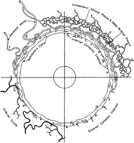
| Fig. 29. Diagrammatic representation of the distal portion of the aqueous outflow pathways from Schlemm's canal. External collector channels (lower right), deep and intrascleral plexuses (upper right), and aqueous veins (1 and 2) (upper left). (From Hogan M, Alvarado J and Weddell J. Histology of the Human Eye. Philadelphia: WB Saunders, 1971.) |