|
|
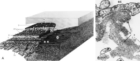
| Fig. 25. A. Anterior ciliary muscle tendons (T) and their connections with the trabecular meshwork. Tendons from the longitudinal bundle of the ciliary muscle (CM) extend to the scleral spur (SP), into the outermost corneoscleral trabeculae, and into the juxtacanalicular region contributing to the cribriform plexus. Connecting fibrils (CF) extend from the plexus toward the endothelial cells (E) lining the inner wall of Schlemm's canal (Sc). B. Immunoelectron micrograph shows a single connecting fibril (c) attaching to the endothelium (E) of the inner wall of Schlemm's canal (SC). Scattered small black dots represent colloidal gold staining for elastin, confirming that these connecting fibrils contain this protein. (A from Rohen JW. Why is intraocular pressure elevated in chronic simple glaucoma? Anatomical considerations. Ophthalmology 90:758, 1983. B Reproduced with permission of Swets and Zeitlinger Publishers and from Gong H, Trinkaus-Randall V, Freddo T: Ultrastructural immunocytochemical localization of elastin in normal human trabecular meshwork. Curr Eye Res 8:1071, 1989.) |