|
|
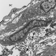
| Fig. 24. Transmission electron micrograph demonstrating the appearance of the connective tissue matrix and cells in the juxtacanalicular region (JCT). The matrix is composed of collagen (c) and elastin and the cells are devoid of a basal lamina. The JCT cells extend mushroom-like cell processes (arrowheads) that attach to endothelial cells (E) of the inner wall of Schlemm's canal (SC). (From Freddo T: Ocular anatomy and physiology related to aqueous production and outflow. In: Fingeret M, Lewis T (eds). Primary Care of the Glaucomas. 2nd ed. New York: McGraw-Hill, 2001.) |