|
|
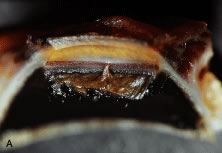
| Fig. 19. Macroscopic photograph (a) of angle structures viewed from the perspective shown in the corresponding sketch (b). Schlemm's canal is filled with blood in this specimen, demonstrating its relationship to the other angle structures. An iris process is also shown. (From Freddo T: Ocular anatomy and physiology related to aqueous production and outflow. In: Fingeret M, Lewis T (eds). Primary Care of the Glaucomas. 2nd ed. New York: McGraw Hill, 2001.) |