|
|
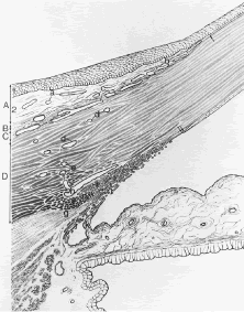
| Fig. 17. Drawing of the limbus to illustrate the structures evident by light microscopy. The limbal conjunctiva (A) is formed by an epithelium (1) and an areolar connective tissue stroma (2). Tenon's capsule (B) forms an ill-defined connective tissue layer of the episclera (C). The limbal corneosclera occupies area D. a, conjunctival vessels; b, corneal arcades; single arrow, terminus of Bowman's layer. The triangular tissue wedge of the trabecular meshwork (I) originates at Schwalbe's line (double arrows) and broadens posteriorly to become continuous with the root of the iris and the ciliary body. An iris process (k) extends from the iris surface to the trabecular meshwork. Entering the posterior margin of the trabecular meshwork is the scleral spur (f), to which tendons of the longitudinal bundle of the ciliary muscle (g) are attached. Aqueous passing through the trabecular meshwork enters the canal of Schlemm (h), deep scleral plexus (e), intrascleral plexus (d), and finally the episcleral vessels (c). (From Hogan M, Alvarado J, Weddell J. Histology of the Human Eye. Philadelphia: WB Saunders, 1971.) |