|
|
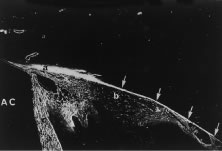
| Fig. 16. Light micrograph demonstrating the distribution of fluoresceinated dextran after intracameral perfusion into the anterior chamber (AC). The tracer has moved posteriorly into the angle tissues (a) and through the ciliary muscle (b) to finally reach the supraciliary and suprachoroidal space (arrows). Tracer has also freely permeated the iris stroma (c). (From Tripathi R. Uveoscleral drainage of aqueous humor. Exp Eye Res 1977;25(Suppl):305.) |