|
|
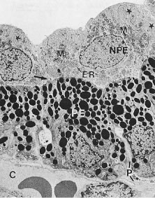
| Fig. 11. Electron micrograph of the ciliary epithelium (cynomolgus monkey, pars plicata). The posterior chamber is at the top and the ciliary body stroma, with its fenestrated capillaries (C), is at the bottom. Extensive basolateral infoldings of the nonpigmented ciliary epithelial cells (NPE) are evident (stars). The apical surfaces of both layers face one another (arrow). PE, Pigmented ciliary epithelium; M, mitochondria; ER, endoplasmic reticulum; P, paracellular space. (Lütjen-Drecoll E: Functional morphology of the ciliary epithelium. In: Lütjen-Drecoll E (ed): Basic Aspects of Glaucoma Research. Stuttgart, Germany: FK Schattauer, 1982, p. 79.) |