|
|
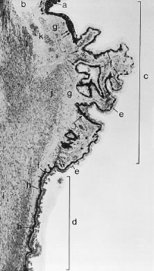
| Fig. 4. Light micrograph of ciliary body of a 50-year-old man. The iris root (a) and the anterior chamber angle (b) are seen at the top. The pars plicata (c) and a portion of the pars plana (d) are included within brackets; they are lined by the nonpigmented (e) and pigmented (f) ciliary epithelium. The basement membrane of the pigmented ciliary epithelium increases (h) in thickness with age. The ciliary body stroma (g) and the three portions of the ciliary muscle (i, longitudinal; j, radial; k, circular bundle) are also shown. (From Hogan M, Alvarado J, Wedell J: Ciliary body and posterior chamber. In: Histology of the Human Eye. Philadelphia: WB Saunders, 1971, p 270.) |