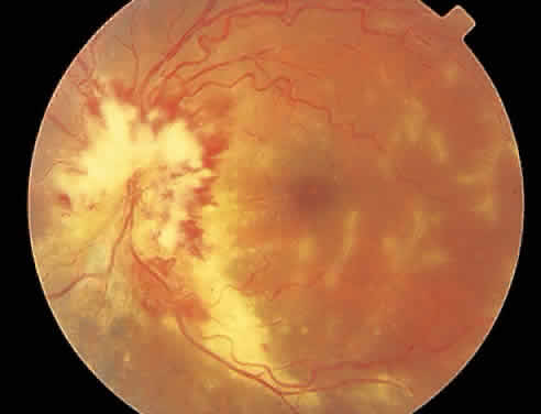
Fig. 2.
Active cytomegalovirus retinitis concentrated around the optic nerve. Temporal to the fovea, there is perivascular whitening with a frosted branch angiitis appearance.