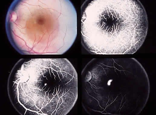
Fig. 32.
Color photograph shows serous detachment of the macula. Early in the angiogram, a hyperfluorescent spot is noted nasal to the fovea. This enlarges on the subsequent frames.