|
|
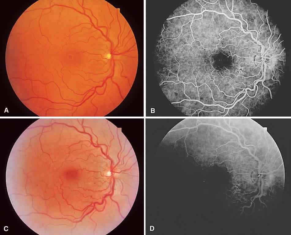
| Fig. 31. A & B. Preoperative photograph shows a stage 3 macular hole with focal hyperfluorescence seen angiographically. C & D. Postoperative photograph shows that the hole is closed and the fovea is flat. Angiogram shows resolution of the hyperfluorescence. |