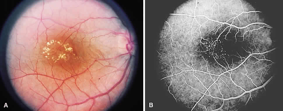
Fig. 25.
A.
Group 1B idiopathic juxtafoveolar retinal telangiectasia. Circinate hard exudate and edema are noted.
B.
Angiography shows telangiectatic change in the area of the exudate.