|
|
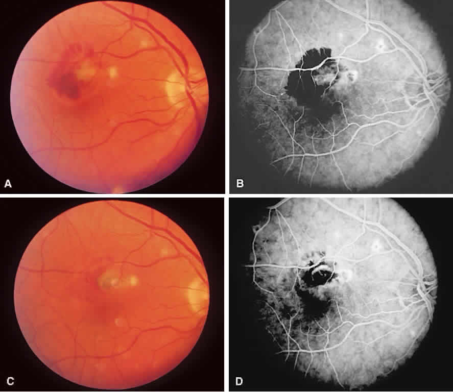
| Fig. 15. A. Histo spots are noted in association with choroidal neovascularization (CNV) membrane (dirty green) and subretinal hemorrhage. B. Corresponding angiogram shows transmitted fluorescence from histo spots, mild peripapillary atrophy, blocked fluorescence from subretinal hemorrhage, and CNV in association with a histo spot. C. After laser treatment to the CNV complex, hemorrhage has resolved and there is early scarring of the CNV membrane. D. Corresponding angiogram shows staining of the scar. |