|
|
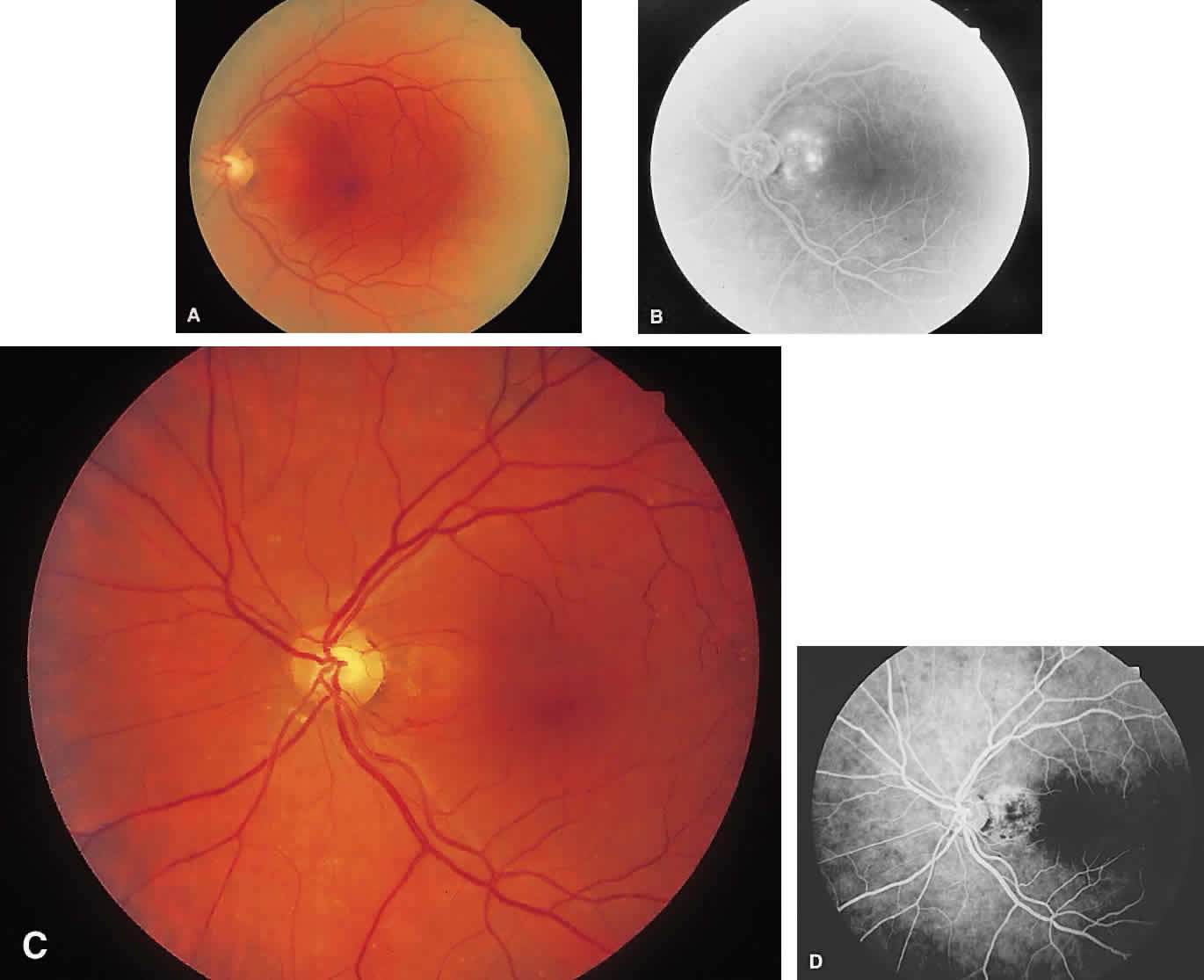
| Fig. 13. A. Neurosensory retinal detachment is noted extending from the optic nerve to the fovea. B. Angiogram shows hyperfluorescence near the temporal border of the nerve from a peripapillary choroidal neovascularization membrane. Note how the margins of the detachment are made evident by the dye. This cushion of fluid protects the neurosensory retina as the choroidal neovascularization membrane is treated with laser photocoagulation. C. Two weeks after laser treatment, a laser scar can be seen adjacent to the nerve. The neurosensory detachment has resolved. D. Posttreatment angiogram shows staining of scar. No detachment is noted. |