|
|
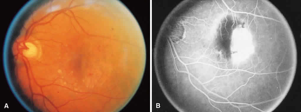
| Fig. 10. A. Atrophy temporal to the fovea and rolled pigment epithelium centrally, created by a retinal pigment epithelial tear in the left eye. B. Fluorescein angiogram showing intense hyperfluorescence created by the window defect after a retinal pigment epithelial tear. The hypofluorescence corresponds to the area where the pigment epithelium rolled together in accordion fashion. |