|
|
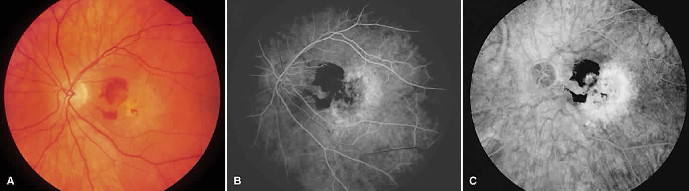
| Fig. 9. A. Color photograph showing subretinal hemorrhage associated with drusen and pigment change. B. Early-phase angiogram shows blocking from subretinal hemorrhage (note overlying retinal vessels) associated with early, diffuse macular hyperfluorescence. The lesion is occult as no classic subretinal neovascularization is noted. C. Late-phase angiogram shows further leakage. |