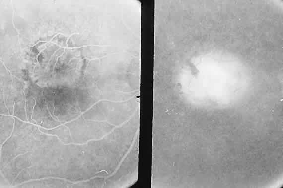
Fig. 8.
Angiogram shows classic subretinal neovascularization with lacy hyperfluorescence and diffuse leakage later from the membrane.