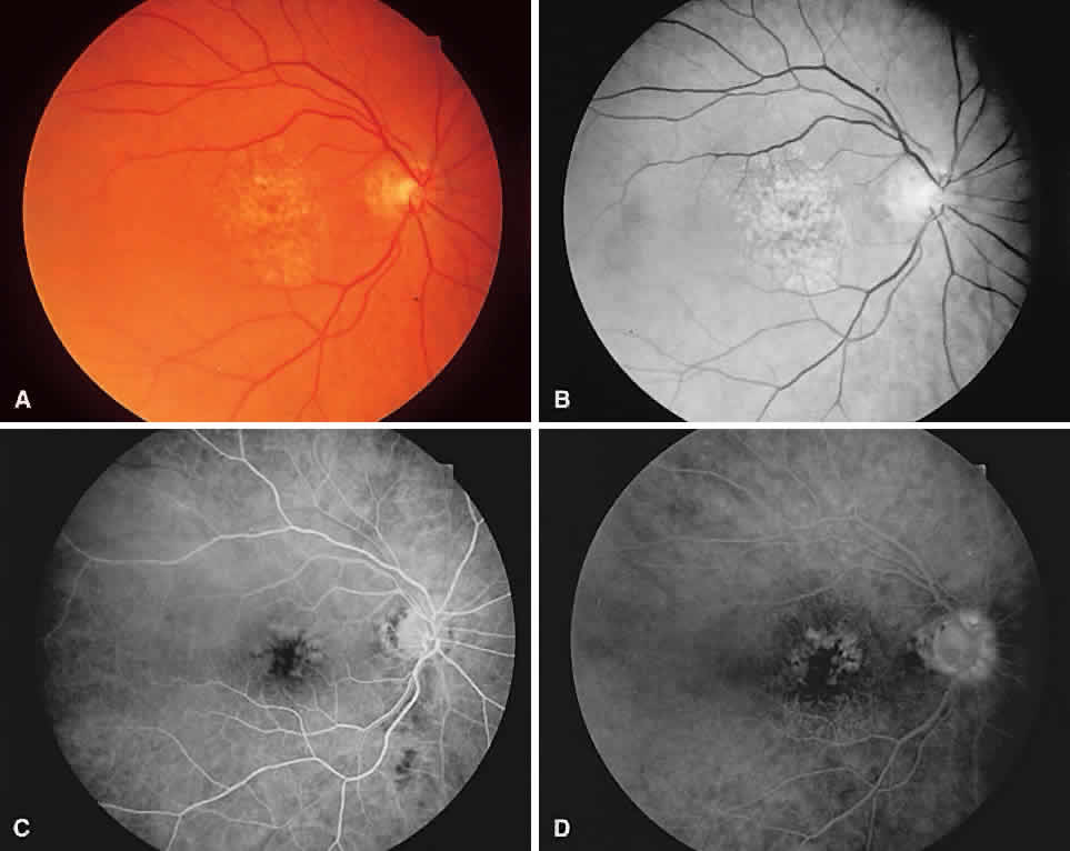
Fig. 3.
Color (
A
) and red-free (
B
) photgraphs of a fundus with soft drusen and hyperpigmentation. Soft drusen hyperfluoresce during the early phase of angiography (
C
) and stain in the late phase (
D
).