|
|
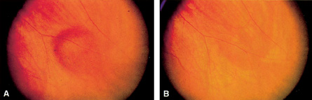
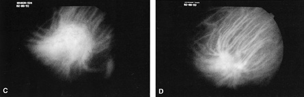
| Fig. 16. A. Gaze-evoked prominence of a vortex vein varix at the inferonasal equator. B. The vortex vein varix disappears in primary gaze. C. ICG angiography shows early filling of the vortex vein varix at 40 seconds. D. ICG hyperfluorescence of the vortex vein varix is decreased in primary gaze. (A to D courtesy of Jerry A. Shields, MD, and Carol L. Shields, MD) |