|
|
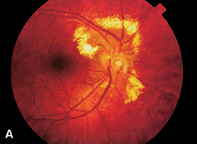
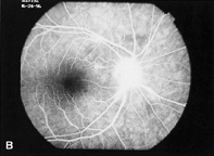
| Fig. 15. A. Fundus photograph of IRVAN demonstrates aneurysms and apparent vascular elongation at the optic nerve with secondary exudation, B. JVFA of IRVAN reveals late staining of the optic nerve head and vasculitis. (A, courtesy of Carmen A, Puliafito, MD) |