|
|
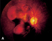
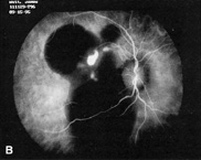
| Fig. 14. A. Small orange-red subretinal lesions in idiopathic polypoidal choroidal vasculopathy are partially obscured by adjacent subretinal hemorrhage. B. ICG hyperfluorescence demonstrates the polypoidal choroidal vascular network, which is contrasted with the hypofluoerscent subretinal hemorrage. (A, courtesy of James J. Augsburger, MD) |