|
|
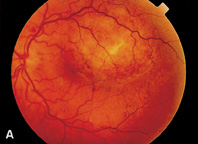
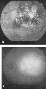
| Fig. 13. A. Circumscribed choroidal hemangioma with fibrous metaplasia on its surface. B. IVFA during arterial phase. Notice the large, early filling of choroidal spaces within the tumor, producing a mottled appearance. C. ICG shows diffuse hyperfluorescence of the circumscribed choroidal hemangioma. This is followed by wash out of ICG dye in late phase angiogram. |