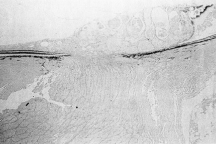
Fig. 12.
Histopathologic study of a retinal cavernous hemangioma of the optic disc. Notice multiple, small, thin-walled, blood-filled spaces. (Courtesy of the Armed Forces Institute of Pathology, Washington, DC)