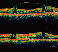
Fig. 7.
Optical coherence tomography. Vitreofoveal traction is visible in the two upper images. The two lower images show full-thickness macular holes of varying sizes. Note the subretinal fluid visible in the lower right image.