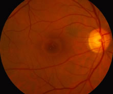
Fig. 6.
Stage 3 macular hole. Note the yellow clumps of presumed glial cells in the base of the macular hole and the cuff of subretinal fluid. In the presence of a complete posterior vitreous separation, this would be classified as a stage 4 hole.