|
|
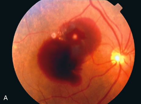
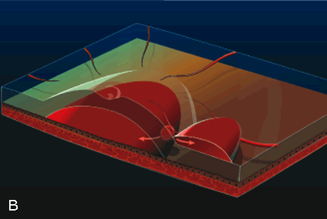
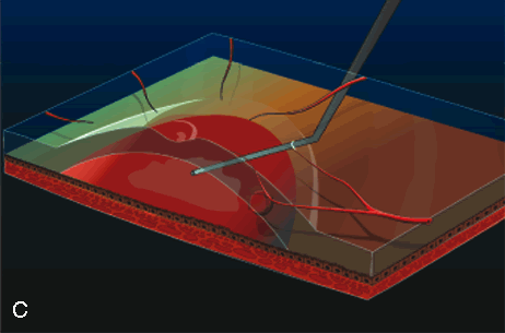
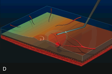
| Fig. 7. A. Color fundus photograph of a macroaneurysm causing subretinal and sub-ILM hemorrhage involving the macula. B. Illustration of the spatial relationship between the submacular blood and sub-ILM blood. C. During vitrectomy surgery, a subretinal cannula is used to inject tissue plasminogen activator into the blood clot. D. After the blood is allowed to lyse over the course of 40 minutes, it is removed using a subretinal cannula. |