|
|
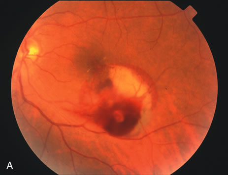
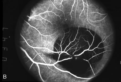
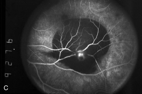
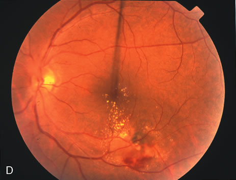
| Fig. 3. A. Color fundus photograph of a macroaneurysm along the inferotemporal arcade with surrounding intraretinal and subretinal hemorrhage. There is retinal edema, hard exudates, and thin subretinal hemorrhage extending into the macula. B. Midphase fluorescein angiography reveals hemorrhage that extends just into the foveal avascular zone, threatening foveal vision. C. Late-phase angiogram reveals staining of the macroaneurysm. D. Color fundus photograph of the same macroaneurysm 6 weeks after laser photocoagulation, demonstrating resolution of the edema and hemorrhage. |