|
|
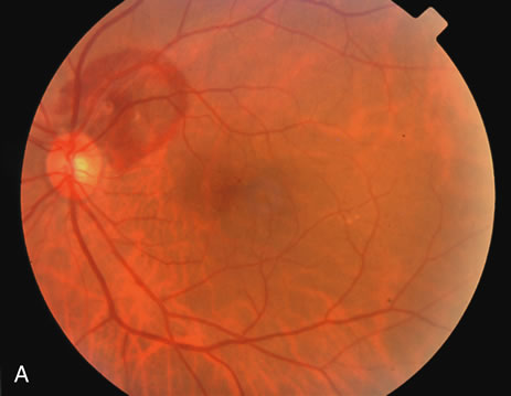
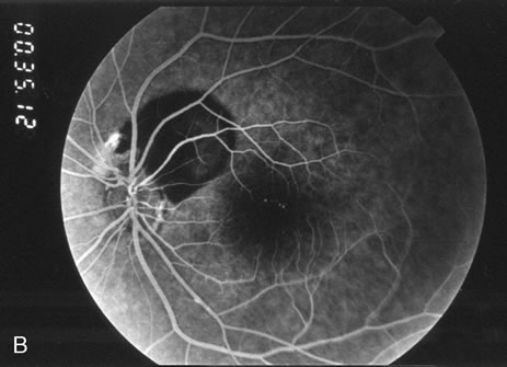
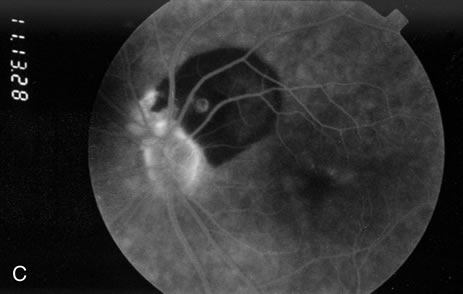
| Fig. 2. A. Color fundus photograph of a 200-micron macroaneurysm that arises from a retinal artery near the optic disc with thin subretinal hemorrhage that does not threaten the fovea. B. The midphase fluorescein angiogram reveals blockage of choroidal but not retinal vascular hyperfluorescence. C. The late-phase angiogram demonstrates staining of the macroaneurysm.[pa[et[ol0] |