|
|
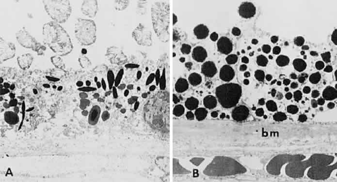
| Fig. 19. A. Electron microscopic findings of normal-appearing retinal pigment epithelium (RPE) granules. B. Electron microscopy within area of congenital hypertrophy of the RPE shows enlarged pigment granules and a thickened basement membrane (bm) of the RPE cells. (Buettner H: Congenital hypertrophy of the retinal pigment epithelium. Am J Ophthalmol 79:177, 1975) |