|
|
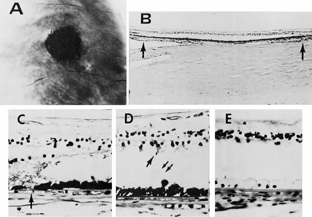
| Fig. 18. A. Fundus photograph of congenital hypertrophy of the retinal pigment epithelium (RPE) without yellow-orange lacunae. B. Histologic preparation shows absence of photoreceptors in an area overlying darker RPE. The region between the arrows corresponds to the lesion shown in A. C. Transition (arrow) from normal RPE on the left to hypertrophied RPE on the right. D. A photoreceptor remnant is shown (single arrow), and periodic acid-Schiff-positive material (double arrows) is seen in the subretinal space. E. Bleached section in the area of hypertrophy reveals a single layer of RPE cells and a thickened Bruch's membrane. (Buettner H: Congenital hypertrophy of the retinal pigment epithelium. Am J Ophthalmol 79:177, 1975) |