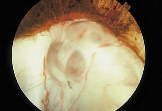
Fig. 13.
Retinochoroidal coloboma involving the optic disc and inferonasal fundus. The borders of the abnormality are nonpigmented, and the defect appears to be filled with fibroglial tissue.