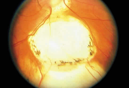
Fig. 12.
Isolated retinochoroidal coloboma with pigmented borders positioned inferior to the nerve head. The sclera is visible through the thinned, overlying retinal tissue.