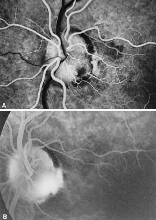
Fig. 9.
A.
Fluorescein angiogram in the laminar venous filling phase of an eye with an inferotemporal optic pit. The pit is hypofluorescent at this time.
B.
Ten minutes after the injection, the pit is hyperfluorescent.