|
|
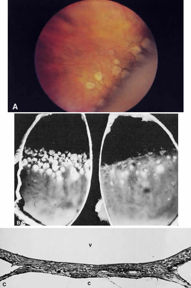
| Fig. 62. A. Paving stones just anterior to the ora serrata. (William Benson: Wills Eye Atlas of Ophthalmology, Fig. 4-58. Philadelphia, Lippincott-Raven, 1996) B. Photograph of the calottes of an autopsy eye showing paving stone degeneration. C. Photomicrograph of paving stone degeneration. The vitreous (v) and choroid (c) are marked for orientation. The retina is detached by an artifact, except in the zone of the paving stone lesion. Notice that the photoreceptors are present to either side of the lesion but not within the lesion. The pigment epithelium also is absent in the area of the lesion. The ophthalmoscopist therefore looks through transparent retina onto choroid and sclera, which accounts for the color of the paving stone lesion. |