|
|
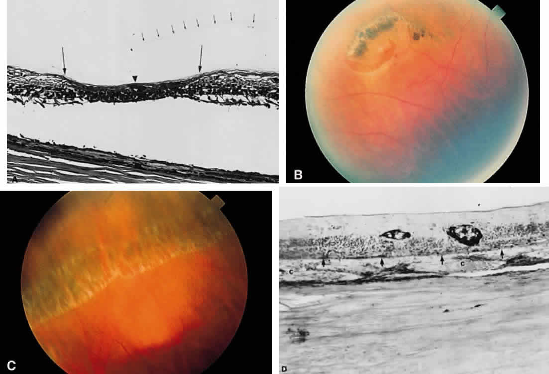
| Fig. 61. (continued) B. Small retinal tear posterior to an area of lattice degeneration. C. Clinical photo of an area of circumferentially oriented lattice degeneration. D. Photomicrograph of primary retinitis pigmentosa. The arrows mark the plane of Bruch's membrane. The photoreceptor elements are missing. Pigment is accumulated around retinal vessels (c, choroid). (B and C, courtesy of William Tasman, MD, Philadelphia, PA). |