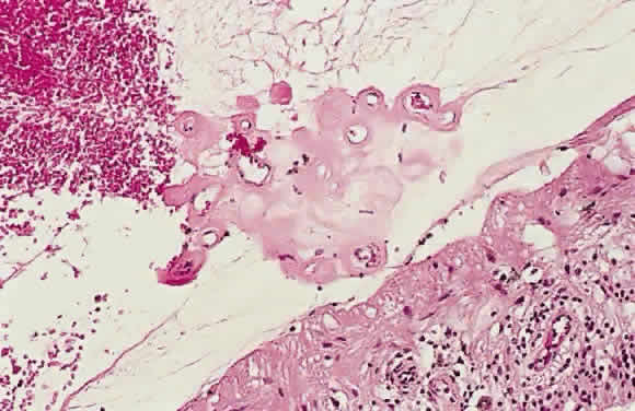
Fig. 54.
Photomicrograph of retinal neovascularization. A tuft of new vessels is seen anterior to the plane of the internal limiting membrane. The detached vitreous contains old hemorrhage. (Courtesy of Ralph C. Eagle Jr, MD, Philadelphia, PA)