|
|
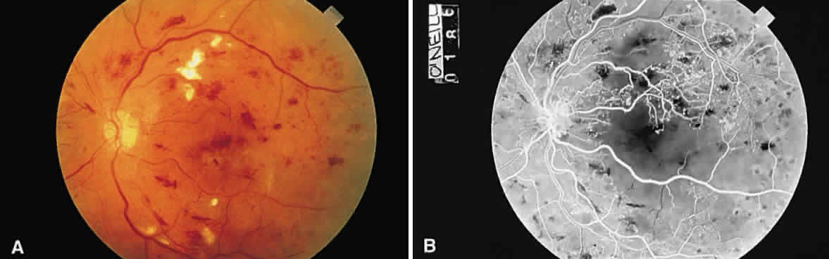
| Fig. 52. A. Fundus photograph of nonproliferative diabetic retinopathy. Notice the dilated veins, the small exudate clusters, the ischemic infarct (cotton-wool spot), and the numerous microaneurysms and small hemorrhages. B. Mid-venous phase fluorescein angiogram of the same eye. Notice the focal area of nonperfusion in the superior temporal arcade (ischemic areas), the fluorescing microaneurysms, and the hemorrhages, which appear dark in contrast to the background fluorescence. (Courtesy of William Tasman, MD, Philadelphia, PA) |