|
|
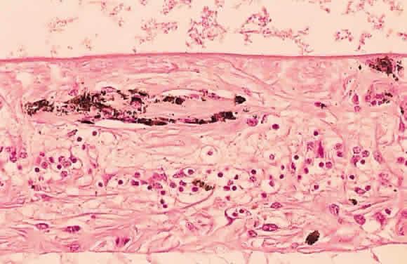
| Fig. 50. Photomicrograph of primary retinitis pigmentosa demonstrating pigment accumulation surrounding a superficial blood vessel and absence of the rods and cones in the posterior retinal layers. (Courtesy of Ralph C. Eagle Jr, MD, Philadelphia, PA) |