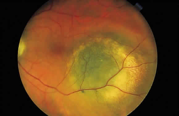
Fig. 48.
Fundus photograph of choroidal melanocytic lesion. Notice its slate gray color, which differentiates it from the jet black congenital hypertrophy of the pigment epithelium.