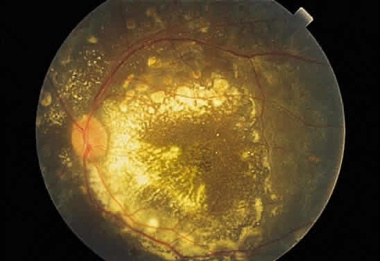
Fig. 43.
Fundus photograph of Coats' disease. Dense exudate is present in the posterior pole. The peripheral lesions, the source of the exudates, are not visible in the photograph.