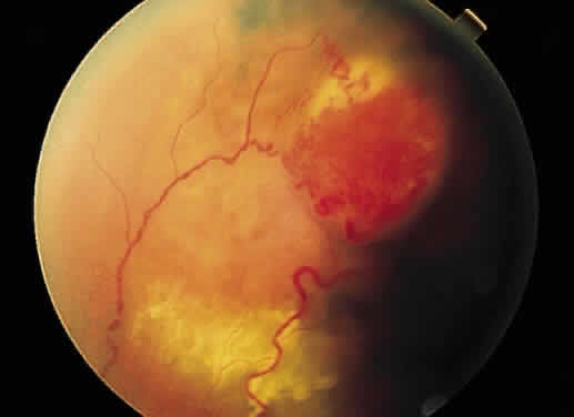
Fig. 42.
Fundus photograph of Von Hippel lesion. Notice the dilated, tortuous vessels leading to and from the tumor and the exudate just visible in the macular region at the bottom of the photo.