|
|
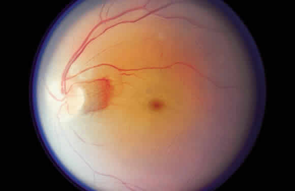
| Fig. 35. Acute central retinal artery occlusion demonstrating generalized opacification of the retina in the posterior pole except for the “cherry-red spot” fovea and a small area of “normal” retina adjacent to the disc supplied by a small cilioretinal artery. |