|
|
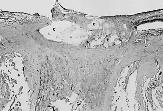
| Fig. 28. Photomicrograph of drusen of the optic nerve head. Calcium (c) is deposited in the nerve anterior to the lamina scleralis. The optic nerve in this case is markedly atrophic. The retina to the left of the nerve is artifactually detached and missing from the plane of the section. |