|
|
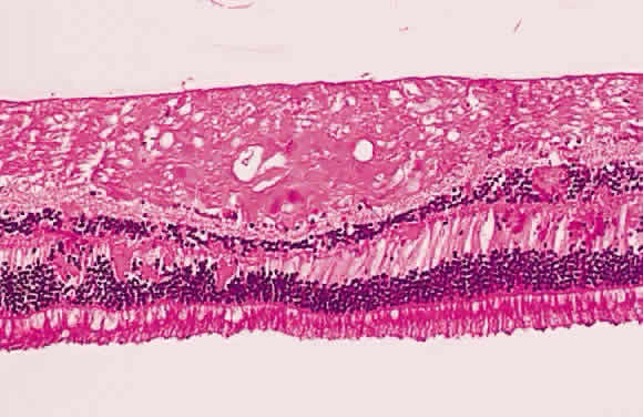
| Fig. 22. Histologic section demonstrating retinal nerve fiber layer infarct. The red dots within the infarct zone are bulbous swellings of nerve fiber axons called “cytoid (cell-like) bodies.” The entire infarcted zone is the histologic counterpart of the clinical cotton-wool spot. (Courtesy of Ralph C. Eagle Jr, MD, Philadelphia, PA) |